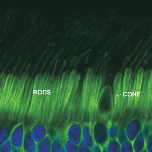
To understand how color blindness works, you first must understand the components of the eye which combine to provide the images you see. You may be familiar with parts such as the retina, iris, lens, cornea etc. The latter parts combine to focus and project light waves onto the retina. Dysfunction in the cornea results in short sightedness or long sightedness etc; however the cause for color blindness lies in the retina.
The retina is responsible for passing any light information that arrives at it down the optic nerve to the brain. The retina is comprised of both ‘rod’ and ‘cone’ cells. The rod cells are extremely sensitive to light, in-fact more than 100x as sensitive as the cone cells. Rod cells become active in low light conditions and usually in the peripheral vision. A simple demonstration of this is to go outside on your next cloud-free night and look at the stars. If you look directly at them, you may not see much, but if you try to study your peripheral vision, you’ll find that much more light is detected, as this is where the rod cells function. However, rod cells have nothing to do with whether or not a person is color blind, all the action happens with the cone cells!
Cone Cells and Color Blindness

“Color” is determined by the wavelength of a stream of light, by detecting the wavelength of incoming light, the eye can determine what color it is looking at. The (normal) eye contains 3 types of cone cells, each containing a different pigment:
- The L-cone detecting long wavelength light (peaking in the yellows – but also responsible for reds).
- The M-cone detecting medium wavelength light (peaking in the greens).
- The S-cone which detects short wavelength light (peaking with blue).
Your brain determines what color it is seeing by observing the ratio between the signals it receives from each of the three types of cones. Color blindness occurs when one or more types of cones are either totally absent, or has a limited spectral sensitivity. By far the most common is congenital (hereditary) red green color blindness, meaning the L-cones and/or M-cones are either damaged or absent. Color blindness is best described by etiology (medical: why things happen) as below, ordered in approximate order of commonality.
Anomalous Trichromacy
Anomalous trichromacy is most the common color vision deficiency with the sub-classification “Deuteranomaly” the most common form of all, found in approximately 5% of all males. Anomoalous trichromacy occurs when one of the three cone pigments is altered, resulting in an impaired sense of color, rather than a total loss. Trichromacy refers to normal three-dimensional color vision – where each type of cone represents one dimension of color. Sub-classifications of anomalous trichromacy:
- Protanomaly is caused by defective L-cones, lowering sensitivity to red hues. To the color blind person suffering protanomaly, this represents itself as a weakened ability to distinguish between some hues of red and green. Protanomaly is often passed from the mother to her son, and is present in 1% of all males. Red green color blindness is the common terminology for this form.
- Deuteranomaly is similarly caused by defective M-cones and is by far the most common form of color blindness, weakening the ability to differentiate red and green hues in as much as 5% of all males. Deuteranomaly, like protanomaly, is also hereditary and typically passed from mother to son, and also known as red green color blindness.
- Tritanomaly is a rare form of color blindness, resulting from weakened S-cones. Tritanomaly affects the ability to discriminate between blue and yellow hues, and is also hereditary. Tritanomaly is more commonly known as blue yellow color blindness.
Dichromacy
Dichromacy, like the three forms of anomalous trichromacy, affects one of the three cones. However in this case, the cone is either absent or non functional. Dichromacy is moderately severe, reducing the color vision of someone with this form of color blindness to two dimensions.
- Protanopia is considered a severe color vision defect and is caused by the total absence of L-Cones. It is the form of dichromacy in which red appears dark, affecting 1% of all males, and is also hereditary.
- Deuteranopia is found when all M-cones are absent, giving a moderate inability to discriminate red – green hues. Like protanopia, it is a form of color blindness where only two types of cone pigments are present, is hereditary, and present in 1% of males. Deuteranopia is also better known as red-green color blindness, but on a somewhat stronger level than protanomaly or deuteranomaly.
- Tritanopia is extremely rare, resulting from a total absence of S-cones, and often labelled blue-yellow color blindness. However, tritanopia could be more aptly named blue green color blindness as people suffering this form see blue hues as greens, and (less commonly) confuse shades of yellow with shades of violet.
Monochromacy
Monochromacy, more commonly referred to as “total color blindness”, is caused by the total absence of either 2 or 3 of the pigmented retinal cones, reducing vision to one dimension. It comes in two forms:
- Rod monochromacy is a rare, non progressive inability to distinguish any color, resulting from non-functioning or absent retinal cones. Rod monochromacy is typically associated with sensitivity to light (Photophobia) and poor vision.
- Cone monochromacy is also a rare type of total color blindness, however is accompanied by relatively normal vision.
Acquired
Color blindness can sometimes be caused through various accidents which result in trauma or damage to the eye. It is also considered possible that long term alcoholism may have an effect, and additionally as a result of many congenital diseases such as diabetes. This information is not so much scientific as observational, and more information on acquired color blindness can be found on the color blindness causes page.